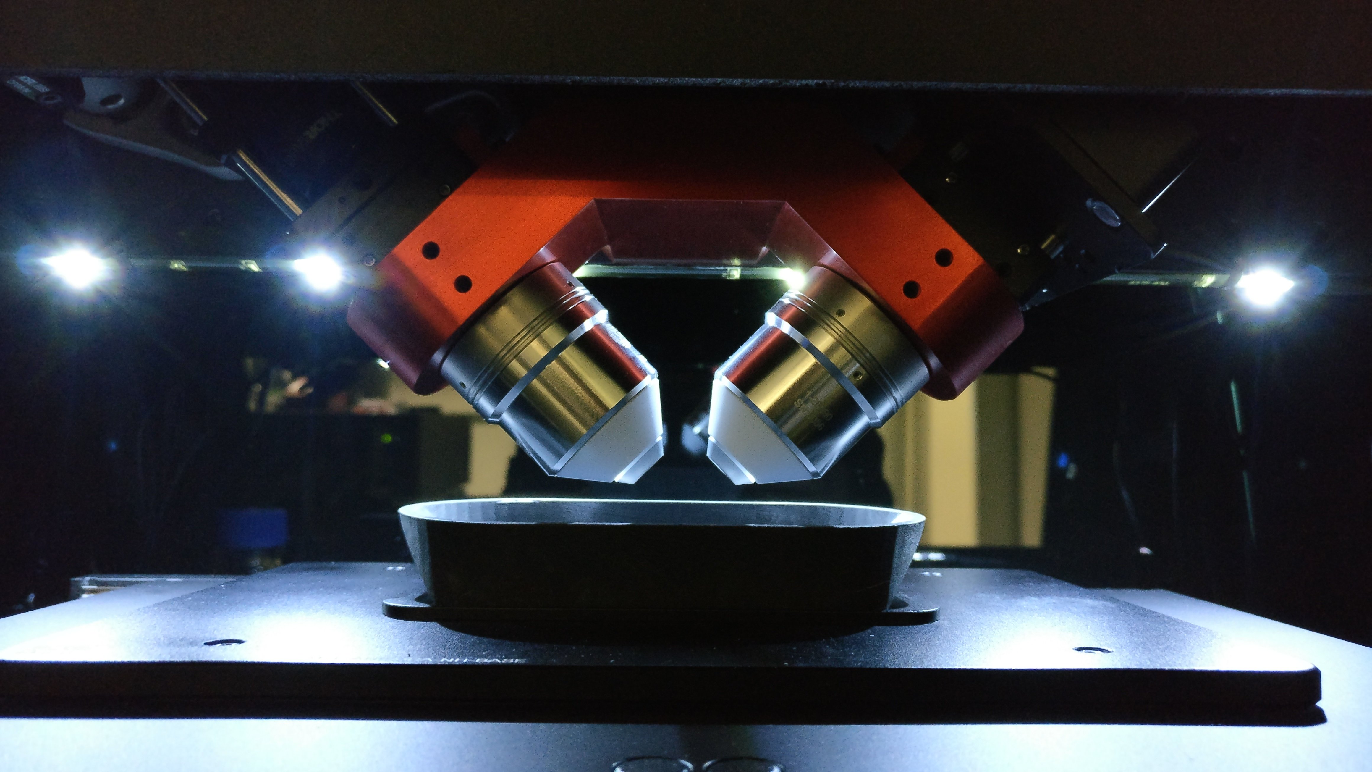
MSquared Aurora Airy Beam
About this microscope
Here you’ll find the hardware and capabilities currently recorded for this microscope. If you are planning an experiment, AIC Turku staff can help with access, training, and configuration choices.
- Light sheet: Light sheet
Software
-
M Squared Cubes Acquisition
Acquisition
Role: Acquisition
Version: Not documented -
M Squared Cubes Deconvolution
Processing
Role: Processing
Version: Not documented
Hardware
Environmental control
- Environmental Control
controlled temperature
Temperature control: Yes
Light sources
-
488 nm Laser Coherent OBIS 488 nm laser
Coherent OBIS
Type: Laser
Wavelength:488 nm
Power:100 mW -
561 nm Laser Coherent OBIS 561 nm laser
Coherent OBIS
Type: Laser
Wavelength:561 nm
Power:80 mW -
640 nm Laser Coherent OBIS 640 nm laser (LX)
Coherent OBIS
Type: Laser
Wavelength:640 nm
Power:75 mW
Notes: FIBER PIGTAIL:UFC: Galaxy
Scanner
- Tandem Scanner (Galvo/Resonant)
Type: Tandem Scanner (Galvo/Resonant)
Light-sheet type: airy_beam
Line rate:8000 Hz
Notes: Galvanometric scanner and remote focusing lenses on illumination/detection; integrated Resonant scanner for beam scanning.
Detectors
- Hamamatsu ORCA-Flash4.0 V3 (C11440-22CU)
Scientific CMOS
Type: Scientific CMOS
Pixel pitch:6.5 µm
Sensor format:2048 x 2048
QE peak:82%
Notes: Supplied with water chiller for camera chip cooling
Objectives
View this microscope's objectives in the catalogue
- 54-10-12 Airy beam dipping objective 17x/0.4 MULTI-IMMERSION
Special Optics
Model: 54-10-12 Airy beam dipping objective
Magnification / NA:17x/0.4
Immersion: Multi-Immersion
Correction: Fluorite
Working distance:12 mm
Product code:54-10-12
AFC compatible: No
Installed: Yes
Specialties: cleared_tissue_imaging
Notes: Excitation and detection objectives designed for dipping media with refractive index range 1.33-1.56 (NA 0.37-0.43), magnification range 15.3x-17.9x across RI. FOV 1.2 mm diameter.
Optical-path elements
- Emission Wheel
Emission
Element type: Filter Wheel
Pos 1: GFP filter — emission 520/40 nm
Pos 2: GFP longpass
Pos 3: RFP longpass
Latest QC measurements
Laser performance
No recent laser-performance measurements are recorded.
Optical resolution (PSF and alignment)
No recent PSF or chromatic-shift measurements are recorded for these objectives.
Objectives without recent measurements
- 54-10-12 Airy beam dipping objective 17x/0.4 MULTI-IMMERSION (
17x_multi)
Information still to confirm
Some details in the facility record are incomplete. If any of these matter for your experiment, please check them with AIC Turku staff.
-
Details needed for this configuration:
-
Software version (
software[].version)