ONI Nanoimager
About this microscope
Here you’ll find the hardware and capabilities currently recorded for this microscope. If you are planning an experiment, AIC Turku staff can help with access, training, and configuration choices.
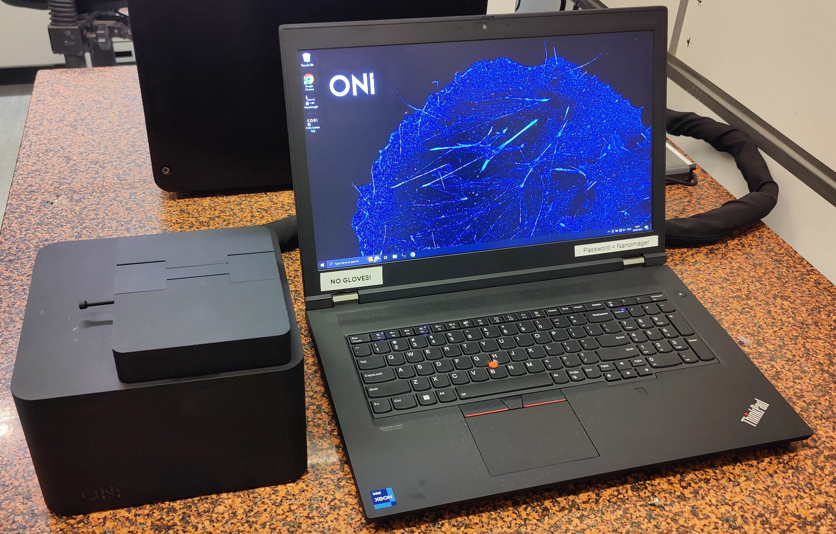
- Widefield fluorescence: TIRF, SMLM
Software
-
NimOS microscope control software
Acquisition
Role: Acquisition
Version: Not documented -
CODI analysis software
Processing
Role: Processing
Version: Not documented
Hardware
Environmental control
- Environmental Control
controlled temperature
Temperature control: Yes
Notes: Resistive heating without active cooling. Suitable for live cell imaging at 37°C.
Light sources
-
405 nm Laser ONI 405 nm violet
ONI
Type: Laser
Technology: diode
Wavelength:405 nm
Power:70 mW
Notes: Power measured at the objective. -
488 nm Laser ONI 488 nm blue
ONI
Type: Laser
Technology: diode
Wavelength:488 nm
Power:250 mW
Notes: Power measured at the objective. -
561 nm Laser ONI 561 nm green
ONI
Type: Laser
Technology: dpss
Wavelength:561 nm
Power:250 mW
Notes: Power measured at the objective. DPSS technology inherently takes ~30 sec to stabilize. -
640 nm Laser ONI 640 nm red
ONI
Type: Laser
Technology: diode
Wavelength:640 nm
Power:220 mW
Notes: Power measured at the objective.
Detectors
- Hamamatsu ORCA-Flash4.0 V3
Scientific CMOS
Type: Scientific CMOS
Pixel pitch:6.5 µm
QE peak:82%
Notes: Digital sCMOS camera, 100 fps full frame. Typically operated with 2x2 binning.
Objectives
View this microscope's objectives in the catalogue
- UPLXAPO100XO 100x/1.45 OIL
Olympus
Model: UPLXAPO100XO
Magnification / NA:100x/1.45
Immersion: Oil
Correction: Apochromat
Working distance:0.13 mm
AFC compatible: Yes
Installed: Yes
Magnification changers
- Custom tube lens
ONI
Manufacturer: ONI
Model: Custom tube lens
Magnification:1.1x
Notes: Custom tube lens provides 110x total magnification with the 100x objective. Combined with 2x2 binning, this yields a pixel size of ~117 nm.
Stages and focus drives
- Z Piezo Stage — ONI Closed-loop Piezo XYZ Stage
ONI
Type: Z Piezo Stage
Autofocus and triggering
- Infrared Reflection Autofocus
Installed: Yes
Type: Infrared Reflection Autofocus
Latest QC measurements
Laser performance
No recent laser-performance measurements are recorded.
Optical resolution (PSF and alignment)
No recent PSF or chromatic-shift measurements are recorded for these objectives.
Objectives without recent measurements
- UPLXAPO100XO 100x/1.45 OIL (
100x_oil)
Information still to confirm
Some details in the facility record are incomplete. If any of these matter for your experiment, please check them with AIC Turku staff.
-
Details needed for this configuration:
-
Software version (
software[].version)